
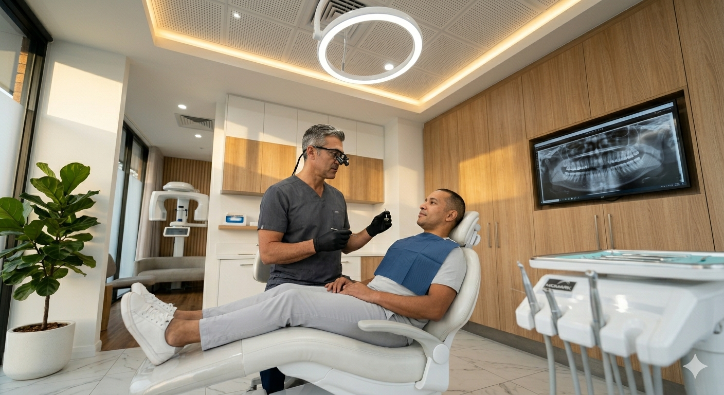
You want clear, safe dental imaging for your child without the fuss. Palmdale clinics use low‑radiation digital x-rays that capture fast, high-quality images so dentists can spot cavities or developmental issues early. These digital x-rays cut exposure and speed up diagnosis, making visits quicker and gentler for kids.
They explain how the technology works in simple terms, show how staff make the experience kid-friendly, and outline when images are needed versus when they’re unnecessary. Expect practical tips on preparing your child, what happens during the appointment, and how follow-up care keeps little smiles healthy.
A local dentist’s focus on comfort and modern safety standards matters, so you know the care aligns with current pediatric guidelines. If you have questions or want to schedule a consultation, the article points toward the next steps to help families in Palmdale feel confident about dental x-rays for kids.
Understanding Digital Dental X-Rays for Children
Digital dental x-rays capture detailed images of a child’s teeth, roots, and jaw with less radiation than older film methods. They help detect cavities between teeth, monitor tooth development, and guide early treatment decisions.
What Makes Digital X-Rays Different
Digital x-rays use an electronic sensor instead of film to record images instantly on a computer screen. Images appear within seconds, allowing the dentist to zoom, adjust contrast, and compare shots quickly during the visit.
Because sensors are more sensitive, exposure time drops by as much as 60–80% compared with traditional film. Many modern pediatric practices in Palmdale use rectangular collimation and fast sensors to further concentrate the beam and reduce scatter.
Digital files are easier to store and share securely with specialists or insurance when needed. This speed and flexibility reduce chair time for children, which helps with cooperation and overall comfort during exams.
Why Digital Imaging Is Safe for Kids
Digital x-rays deliver far lower radiation doses than older film systems, and clinics follow ALARA (As Low As Reasonably Achievable) principles. Protective measures like lead aprons, thyroid collars, and limiting images to what is clinically necessary cut exposure further.
Dentists custom-tailor x-ray schedules for each child based on age, cavity risk, and growth patterns. For example, a low-risk preschooler might need fewer bitewings than an adolescent with active decay.
Regulatory guidelines recommend using digital sensors and modern equipment; most Palmdale pediatric dentists also calibrate machines regularly and use fast exposure settings. Those steps combine to make routine dental imaging a low-risk tool for diagnosis and care planning.
Common Dental Concerns Addressed by X-Rays
Dental x-rays reveal early cavities between teeth that visual exam can miss, especially in the posterior molars. They also show how permanent teeth are developing and whether crowding, missing teeth, or ectopic eruption may require intervention.
X-rays detect problems like root infections, bone loss from periodontal disease, and impacted teeth that could affect orthodontic timing. In trauma cases, imaging helps identify fractures or tooth displacement that need urgent care.
For treatment planning, x-rays guide decisions on fillings, extractions, space maintainers, and orthodontic referrals. Clear images let the dentist explain findings to parents with visual evidence, helping families choose appropriate, timely steps for a child’s dental health.
Benefits of Digital X-Rays in Pediatric Dentistry
Digital x-rays reduce radiation, speed diagnosis, and make visits more comfortable for children and parents. They help dentists spot cavities, monitor jaw growth, and explain findings quickly using images that can be enlarged and shared.
Lower Radiation Exposure
Digital sensors capture images with far less radiation than traditional film. For a typical bitewing series, digital systems can cut exposure by 50–90% depending on sensor type and settings, which matters for children because they are more sensitive to ionizing radiation.
Dentists use lead aprons and thyroid collars in addition to low-dose settings to further protect young patients. Pediatric dental offices follow ALARA (As Low As Reasonably Achievable) principles, adjusting exposure for a child’s size and using only the views needed.
Reducing repeat images also lowers total exposure. Because digital images appear instantly and can be adjusted for contrast, clinicians rarely need retakes for positioning or poor development, keeping cumulative dose down over multiple visits.
Accurate Diagnosis for Growing Smiles
High-resolution digital images reveal early enamel lesions, small interdental cavities, developing permanent teeth, and root formation. This clarity helps dentists make evidence-based decisions about sealants, fillings, or monitoring without waiting for symptoms.
Digital tools allow enlargement, measurement, and side-by-side comparison over time. Tracking tooth eruption patterns and jaw growth on sequential images helps orthodontic planning and early interception when needed.
Images can be emailed or shown on-screen to parents during the visit. Visuals make explanations concrete, increasing parental understanding and consent for recommended preventive or restorative care.
Faster and More Comfortable Visits
Digital x-rays appear on the monitor within seconds, shortening appointment time and reducing the time a child must hold a sensor in their mouth. Faster imaging lowers anxiety and improves cooperation for fearful or fidgety children.
Sensors are thinner and often more flexible than film, which increases comfort during intraoral views. Fewer retakes also mean less time in the chair and fewer stressful moments for both child and caregiver.
Faster results let the dentist discuss findings immediately and show treatment options using the same images. That immediacy supports quick decision-making and clearer communication about next steps.
How Digital X-Rays Work at a Kid-Friendly Practice
Digital dental X‑rays capture clear images quickly while minimizing radiation and keeping kids calm. Staff use short explanations, distraction tools, and child-sized positioning so the process feels fast and safe.
Gentle Techniques for Young Patients
Dental teams use simple, age-appropriate language to explain each step, often demonstrating with a stuffed animal or mirror before taking images. They let the child sit on a parent’s lap when appropriate and offer choices, like which hand to hold a comfort toy with, to give the child control.
Technicians keep exposures brief — usually under a second per image — and take only the views needed for diagnosis. They use bite blocks and soft positioning aids sized for children to ensure stable, comfortable posture and fewer repeat shots.
Staff monitor emotional cues and pause if the child needs a break. They use distraction tools (videos, music, or a tablet) and positive reinforcement, like stickers, immediately after imaging to make the visit pleasant.
Modern Equipment Designed for Safety
Digital sensors replace traditional film, converting X‑ray data to high-resolution images on the clinician’s monitor within seconds. This eliminates chemical processing, speeds diagnosis, and allows immediate review with parents using zoom and annotation tools to explain findings.
Cone-beam and intraoral digital systems used in pediatric settings operate at lower radiation doses than older film systems. Lead aprons with thyroid collars remain standard; the clinic documents exposure levels and follows ALARA (As Low As Reasonably Achievable) principles to minimize dose.
Software enhances image clarity so fewer retakes are necessary, and secure digital records allow teams to compare images over time to track growth and treatment progress. Parents receive clear, annotated images and plain-language explanations to help decide next steps or preventive measures.
Palmdale’s Approach to Family Dental Care
Palmdale dental practices prioritize comfort, prevention, and clear communication for children and parents. They combine kid-friendly exam rooms, digital tools, and appointment flexibility to reduce anxiety and keep care on track.
Personalized Care for Local Families
They tailor preventive plans to each child’s age, cavity risk, and dental history. For toddlers, clinicians focus on early oral hygiene coaching and fluoride varnish. For school-age children, they recommend sealants on permanent molars and schedule digital X-rays only when clinically indicated to limit exposure.
Staff review family dental history, diet, and fluoridation at each visit to adjust recall intervals and home care. They document growth patterns and orthodontic indicators so referrals happen before problems worsen. Bilingual staff and culturally aware education materials help parents follow recommendations accurately.
Insurance and payment options are discussed upfront. Clinics often offer same-week sick slots and extended hours to accommodate school and work schedules. These practical choices reduce missed visits and keep preventive care consistent.
Collaboration Between Parents and Dental Teams
Dentists involve parents in treatment decisions by explaining findings from digital X-rays and visual exams in plain language. They show images on-screen, point out early decay or eruption patterns, and outline specific steps — such as timed brushing, diet changes, or a short sealant procedure.
Hygienists coach parents on at-home techniques using demonstrations and take-home guides. They set measurable goals (e.g., two minutes brushing twice daily, reduced juice to one serving) and schedule follow-ups to track progress. This creates accountability without judgment.
Teams also coordinate with pediatricians and schools when needed, sharing records or care plans to manage issues like thumb-sucking, enamel hypoplasia, or trauma. Parents leave with a clear action list, appointment plan, and contact options for questions.
Preparing Your Child for Their First Digital X-Ray
A calm child, clear expectations, and simple explanations reduce fear and speed the visit. Practical steps include what to say at home, what to bring, and how the team will support the child during the x-ray.
Tips for Easing Anxiety
Parents should explain the procedure in plain words: the x-ray is a quick picture that helps the dentist see teeth inside the mouth. Use specific phrases like, “It will take a few seconds while you sit very still,” to set concrete expectations.
Role-play at home with a flashlight or a small pillow to imitate positioning; this gives the child a sense of control. Bring a favorite toy or blanket to the appointment to provide comfort and a familiar smell.
Ask the clinic ahead if a parent may stand nearby or hold the child’s hand during the exposure. Request that staff use child-friendly language and demonstrate the equipment before starting, which usually calms children faster than abstract reassurances.
Avoid promising the child there will be no discomfort; instead, say the procedure is quick and that the team will help them stay comfortable. Offer praise and a small non-food reward after the visit to reinforce positive behavior.
What to Expect During the Visit
On arrival, staff will confirm medical history, especially recent illnesses or pregnancy exposure in caregivers. They will explain radiation safety: digital x-rays use very low doses and the child will wear a lead apron and thyroid collar for added protection.
The dental assistant will position a small sensor or film inside the child’s mouth or use a handheld digital camera for extraoral images. The child must bite gently on a soft holder for intraoral sensors; this usually takes one or two exposures per area.
Technicians may take images from outside the operatory or step back and press a button; parents often stand behind a barrier or wear a lead apron if allowed. The entire process typically lasts 5–10 minutes in the clinic, with the actual exposures lasting only seconds.
After images are taken, the dentist reviews them on a screen and explains findings in plain terms, showing parents and children any areas of concern or good news. Families should ask about follow-up steps, whether preventive care or monitoring is recommended.
Aftercare and Follow-Up Guidance
The office will explain next steps, expected sensations, and who to call with questions. Parents should track any changes in comfort, tooth appearance, or behavior and keep scheduled follow-ups.
Interpreting X-Ray Results
The dentist reviews digital images with the parent and points out specific findings, such as cavities, unerupted teeth, trauma, or growth patterns. They will show the image on the monitor, label areas of concern, and compare with prior films when available to highlight changes.
If the x-ray shows decay, the team explains treatment options (sealants, fillings, or monitoring) and timelines. If dental development looks atypical, they may recommend an orthodontic consult or periodic radiographs to watch tooth movement. Radiation exposure from modern digital x-rays is very low; the dentist will note the dose and justify the need.
Parents should ask for copies of images or written notes and request clarification about any terms they don’t understand. If the child experiences new pain, swelling, or injury after imaging, contact the practice immediately for reassessment.
Encouraging Healthy Oral Habits at Home
Parents should enforce twice-daily brushing with a fluoride toothpaste appropriate for the child’s age and use a pea-sized amount for children over two. Supervise brushing until the child can reliably spit and perform an effective two-minute routine.
Daily flossing should begin once adjacent teeth contact each other; the dentist will demonstrate proper flossing technique during the visit. Limit sugary snacks and sticky treats; offer water after meals to help clear food particles and reduce acid exposure.
Schedule regular dental check-ups every six months or as recommended after the x-ray findings. Keep a simple log of any tooth pain, sensitivity, or changes in bite to share at follow-ups, and call the office with questions about diet, fluoride, or preventive treatments like sealants.
When Are Digital X-Rays Recommended?
Digital x-rays help detect cavities, check tooth and jaw development, and monitor existing dental work. They use lower radiation than film and provide faster images, which makes them useful in specific, routine situations.
Routine Check-Ups and Preventive Care
Dentists recommend digital bitewing x-rays for children when they expect cavities between teeth or when a child reports pain that can’t be seen visually. For low-caries-risk children, bitewings are often taken every 12–24 months; for higher risk, every 6–12 months. Digital sensors capture clear images quickly, reducing exposure and the need for retakes.
Pediatric dentists also use periapical digital x-rays to evaluate the entire tooth and root when a child has prolonged sensitivity, swelling, or trauma. These images guide treatment decisions like fillings or root canal therapy and help document changes over time. Technicians position sensors to minimize discomfort for smaller mouths.
Evaluating Dental Growth and Development
Orthodontic and growth assessments rely on panoramic and cephalometric digital x-rays to view all developing teeth, jaw relationships, and tooth eruption patterns. Providers schedule these when mixed dentition begins (around ages 6–8) or when early signs of crowding, asymmetry, or delayed eruption appear. Panoramic images reveal unerupted permanent teeth and supernumerary teeth that could affect spacing.
Cephalometric x-rays support treatment planning for braces or growth-modification appliances by showing jaw alignment and facial proportions. Repeat imaging occurs only when clinical findings change or to monitor progress, keeping frequency as low as reasonably achievable. Parents receive clear explanations of why each image matters and how it informs comfort-focused care.
Choosing the Right Dentist in Palmdale for Your Child
Parents should look for a dentist who specializes in pediatric care or has extensive experience treating children. A provider who understands developmental stages and uses child-friendly communication helps reduce anxiety and builds trust.
Check whether the office uses digital X-rays and explains the benefits clearly. Digital X-rays reduce radiation exposure and produce faster images, which makes visits quicker and less stressful for kids.
Observe the clinic environment during a visit or virtual tour. Friendly staff, age-appropriate decor, and distraction tools like books or videos signal a child-centered practice that prioritizes comfort.
Confirm the dentist’s credentials and continuing education in pediatric dentistry and radiography. Licensing and up-to-date training show they follow safety standards and modern techniques.
Ask practical questions before booking an appointment:
- Does the office use digital radiography?
- How do they minimize radiation and explain procedures to children?
- Are emergency appointments available?
Read reviews and request recommendations from local parents or community groups. Firsthand accounts reveal how a practice handles nervous children, preventive care, and follow-up.
Encourage families to schedule an initial consultation to meet the dentist and staff. A short, friendly visit helps determine fit and lets parents observe how the team interacts with their child.
Frequently Asked Questions
These answers explain safety, timing, frequency, and comfort measures for digital dental X-rays for children in Palmdale, CA, and describe how digital imaging improves early detection of cavities and orthodontic needs.
Are digital dental X-rays safe for children, and how much radiation is involved?
Digital dental X-rays use much less radiation than traditional film X-rays. A typical bitewing digital X-ray exposes a child to about 0.5 to 5 microsieverts, roughly equivalent to a few days of natural background radiation.
Pediatric dental offices in Palmdale use lead aprons, thyroid collars, and modern sensors to further reduce exposure. They follow ALARA (As Low As Reasonably Achievable) principles to limit dose while getting the needed diagnostic detail.
At what age should a child start getting dental X-rays?
Dentists usually take the first X-ray when cavities are suspected or when primary teeth have unexplained problems. If the child has routine, low-risk visits and no symptoms, X-rays may be delayed until permanent teeth start to erupt, often around ages 6–7.
If the child has a history of cavities, poor tooth development, or trauma, dentists may take images earlier and more often to guide treatment.
How often does my child actually need X-rays during routine checkups?
Frequency depends on the child’s risk level. For low-risk children, dentists often recommend bitewing X-rays every 12–24 months.
For children with active decay, orthodontic concerns, or developmental issues, X-rays may be taken every 6–12 months. The dentist balances diagnostic benefit against radiation exposure when setting a schedule.
What is the “rule of 7” in pediatric dentistry, and how does it affect X-ray timing?
The “rule of 7” stages dental development in roughly seven-year intervals: 0–7, 7–14, and 14–21, aligned with primary, mixed, and permanent dentition phases. Dentists use these stages to anticipate eruption patterns and plan imaging when teeth are changing.
During the mixed dentition phase (about ages 6–12), X-rays become more useful for monitoring erupting permanent teeth and early orthodontic signs. Timing then focuses on specific developmental milestones rather than arbitrary calendar dates.
How can I help my child feel calm and comfortable during dental X-rays?
Explain the process in simple, positive terms and show the equipment briefly so it seems familiar. Bring a favorite toy or let the child watch a short video when allowed to reduce anxiety.
Dentists and hygienists in Palmdale use child-friendly language, step-by-step guidance, and gentle positioning. They also offer distractions and praise to make the procedure quick and stress-free.
Do digital X-rays help catch cavities and orthodontic issues earlier than a visual exam alone?
Yes. Digital X-rays reveal early interproximal cavities, root issues, and unerupted teeth that a visual exam can miss. They also provide clear images for assessing tooth position and jaw development relevant to early orthodontic decisions.
Digital images can be enhanced, stored, and compared over time, helping the dentist detect subtle changes sooner and plan less invasive, preventive care when possible.